

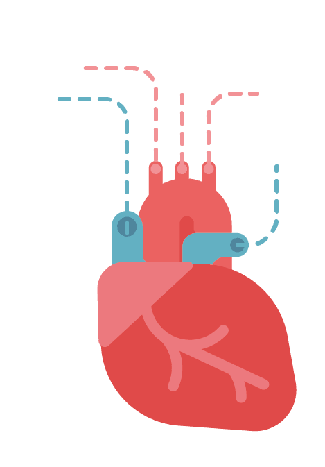
Your heart beats around 100,000 times a day and pumps roughly 2,000 gallons of blood — yet it is only about the size of your fist. Most of us barely notice it working. This guide explains how the heart is built, how it moves blood around your body, and what keeps it running.
How the heart develops
In an early embryo, the heart begins as a simple tube. Over the first weeks of development this tube twists, folds and divides, forming chambers and walls. By around 20 weeks it is essentially fully formed. After birth it changes very little in structure — one reason cancer originating in the heart is extremely rare.
The four chambers
Although it looks like one muscle, the heart is divided into left and right sides, each with an upper chamber (atrium) and a lower chamber (ventricle).
The right side receives deoxygenated blood returning from the body. The right atrium fills, then passes blood through the tricuspid valve into the right ventricle. When the right ventricle contracts, the tricuspid valve snaps shut and blood is pushed through the pulmonary valve into the lungs.
The left side receives freshly oxygenated blood from the lungs. The left atrium fills and feeds blood through the mitral valve into the left ventricle — by far the most muscular chamber. When it contracts, the mitral valve closes and blood surges through the aortic valve into the aorta and out to the entire body.
The four valves act as one-way doors, and the familiar "lub-dub" of a heartbeat is the sound of them closing in sequence.
The electrical system
The heart's coordination comes from a tightly regulated electrical system. The sinoatrial (SA) node in the right atrium acts as the natural pacemaker, generating an impulse that spreads rapidly across both atria, causing them to contract and top up the ventricles.
The impulse then reaches the atrioventricular (AV) node, a checkpoint just above the ventricles, which deliberately delays it by a fraction of a second. That pause is what allows the atria to finish emptying before the ventricles fire.
From there the signal races down the bundle of His, splits into left and right bundle branches, and spreads through the Purkinje fibres, producing a coordinated contraction that squeezes blood upward and outward. The system then recharges and fires again. This is precisely the activity an ECG measures.
The heart's own blood supply
Curiously, the heart cannot use the blood passing through its chambers. It has a dedicated supply: the coronary arteries, which branch off the aorta just above the aortic valve and wrap around the outside of the heart, sending vessels inward to feed every layer of muscle.
This matters enormously. Because the coronary arteries are relatively narrow and the heart never rests, a blockage in one of them starves heart muscle of oxygen very quickly — which is exactly what happens in a heart attack.
What a healthy heart looks like
- Resting heart rate: typically 60–100 beats per minute in adults; fit people are often lower
- Blood pressure: generally below 120/80 mmHg is considered ideal
- Ejection fraction: the proportion of blood the left ventricle pushes out per beat, normally 50–70%
Looking after it
The fundamentals are unglamorous but genuinely effective: don't smoke, move regularly, keep blood pressure and cholesterol in range, limit alcohol, sleep properly, and eat a diet built on vegetables, wholegrains, pulses, oily fish and nuts.
Checking in on your heart
Rightangled's home blood tests cover cholesterol, HbA1c and other cardiovascular markers, and our DNA tests look at inherited risk. Results are reviewed by our clinical team, which includes GPhC-registered independent prescribers, with medical oversight from our doctor, Dr Abdullah.
Verify us with the General Pharmaceutical Council (registration 9011933), see our LegitScript certification, and read reviews on Trustpilot.
Related reading: what causes heart disease.
This article is for general information and does not replace personalised medical advice.

Share:
No More ‘One Size Fits All’
Basic Heart Health